Overview
The visual system processes various kinds of visual signals, such as color and motion. The retina in the eyes starts capturing all visual signals, encodes them to distinct neural pathways, and sends them out to the brain. We have conducted in vitro experiments using the mouse retinal tissue to elucidate neural networks in the retina and signal processing.
Signaling Kinetics of ON and OFF Bipolar Cells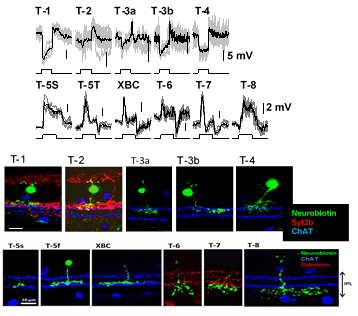
Visual signaling in the retina begins with rod and cone photoreceptors, which provide synaptic inputs to bipolar cells, second-order neurons, then to amacrine and ganglion cells, third-order neurons. Many subtypes of bipolar, amacrine, and ganglion cells have been identified, which are thought to form distinct neural networks and separately encode fundamental components of the visual field, such as color, contrast, or motion. This process is known as parallel processing. In order to understand how individual bipolar cell types transmit different types of information, our lab has performed various studies to examine bipolar cell signaling kinetics.
We examined the signaling kinetics of both ON and OFF bipolar cells using patch clamp physiology to record responses to either flash of light or sine-wave light flicker stimuli. We found that different types of bipolar cells had distinct temporal profiles (Figures). High-frequency tuned cells are more likely to transmit information such as object motion, while low-frequency tuned cells are more likely to transmit static information such as color or contrast.
1. Ichinose T, Fyk-Kolodziej B & Cohn J. (2014). Roles of ON cone bipolar cell subtypes in temporal coding in the mouse retina. J Neurosci 34, 8761-8771.
2. Hellmer CB, Zhou Y, Fyk-Kolodziej B, Hu Z, Ichinose T. Morphological and physiological analysis of type-5 and other bipolar cells in the Mouse Retina. Neuroscience. 2016 Feb 19;315:246-58.
3. Ichinose T, Hellmer CB. Differential signaling and glutamate receptor compositions in the OFF bipolar cell types in the mouse retina. J Physiol. 2016 Feb 15;594(4):883-94.
Dopamine and Bipolar Cell Signaling
The catecholamine neurotransmitter, dopamine, is an important contributor to retinal signaling. Dopamine is released from a unique type of amacrine cell during the daytime in response to light and is not released at night when light is absent. Using transgenic mice where dopamine receptor 1 (D1R) expressing cells contain a red fluorescent protein (RFP) as well as immunohistochemistry, we identified 7 types of bipolar cells that contain D1Rs and 5 types that did not express these receptors. Current electrophysiology experiments in the lab suggest that these receptors alter the signaling kinetics of D1R-expressing bipolar cells in the presence of dopamine (daytime), but this work is still ongoing.
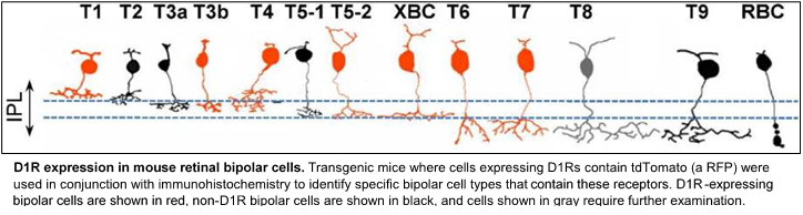
1. Farshi, P., Fyk-Kolodziej, B., Krolewski, D.M., Walker, P.D., & Ichinose, T. (2016) Dopamine D1 receptor expression is bipolar cell type-specific in the mouse retina. Journal of Comparative Neurology 524(10):2059-79.
2. Hellmer, CB., Bohl, JM., Hall, LM., Koehler, CC., & Ichinose, T. (2020) Dopaminergic modulation of signal processing in a subset of retinal bipolar cells.
Alpha-7 nicotinic acetylcholine receptors in a subset of bipolar cells contribute to direction selectivity in Starburst Amacrine cells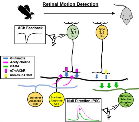 Retinal bipolar cells are second-order neurons that transmit basic features of the visual scene to postsynaptic partners. However, their contribution to motion detection has not been fully appreciated. Here, we demonstrate that cholinergic feedback from starburst amacrine cells (SACs) to certain presynaptic bipolar cells via alpha-7 nicotinic acetylcholine receptors (α7-nAChRs) promotes direction-selective signaling. Patch clamp recordings reveal that distinct bipolar cell types making synapses at proximal SAC dendrites also express α7-nAChRs, producing directionally skewed excitatory inputs. Asymmetric SAC excitation contributes to motion detection in On-Off direction-selective ganglion cells (On-Off DSGCs), predicted by computational modeling of SAC dendrites and supported by patch-clamp recordings from On-Off DSGCs when bipolar cell α7-nAChRs is eliminated pharmacologically or by conditional knockout. Altogether, these results show that cholinergic feedback to bipolar cells enhances direction-selective signaling in postsynaptic SACs and DSGCs, illustrating how bipolar cells provide a scaffold for postsynaptic microcircuits to cooperatively enhance retinal motion detection.
1. Hall, LM., Hellmer CB, Koehler CC, & Ichinose T. (2019) Bipolar cell type-specific expression and conductance of alpha-7 nicotinic acetylcholine receptors in the mouse retina. IOVS Apr 1:60(5):1353-1361.
2. Hellmer CB, Hall LM, Hohl JM, Sharpe ZJ, Smith RG, & Ichinose T. (2021) Cholinergic feedback to bipolar cells contributes to motion detection in the mouse retina. Cell Reports Dec 14:37(11): 110106.